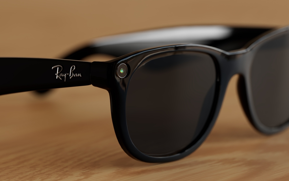
Ray-Ban Meta Glasses
Ray-Ban has long been recognized as a leader in timeless style, but now the brand is taking a bold step into the future. In collaboration with Meta, Ray-Ban has introduced a groundbreaking innovation - Ray-Ban Meta Glasses. These glasses are a fusion of iconic design and next-generation technology that allows you to capture, share, and stay connected like never before.
What Are Ray-Ban Meta Glasses?
Ray-Ban Meta Glasses go beyond vision correction or sun protection - they seamlessly integrate a camera, speakers, and voice-activated AI assistance, allowing users to capture, share, and interact hands-free. With their classic Ray-Ban frames, these glasses look just like traditional eyewear, but they’re packed with cutting-edge features hidden in plain sight.
Key Features and Technology
Built-in Cameras: Capture photos and videos instantly with a simple voice command or tap on the frame. The dual 12MP cameras allow for high-quality media with depth and detail.
Open-Ear Audio: Discreet speakers positioned near your ears provide clear sound without blocking ambient noise, letting you listen to music, take calls, or hear notifications seamlessly.
Meta AI Integration: Access Meta’s AI assistant through voice commands. Ask questions, control your device, or get updates - all without taking your phone out.
Live Streaming and Sharing: Stream directly to social media platforms like Instagram and Facebook in real time, making it easy to share your perspective with your audience.
Touch and Voice Controls: Intuitive touch-sensitive temples and responsive voice commands give you full control over your smart features.
Transitions® Lenses Option: Enjoy lenses that automatically adapt to changing light conditions for optimal comfort both indoors and outdoors.